Yan Luo, Binbin Zhai, Min Li, Wenjingli Zhou, Jinglun Yang, Yuanhong Shu*, and Yu Fang*. J. Colloid Interface Sci., 2024, 660, 513-521.

Wearable SERS substrates have gained substantial attention for health monitoring and other applications. Current designs often rely on conventional polymer substrates, leading to discomfort and complexity due to the need of additional adhesive layers. To address the issues, we fabricate a flexible, uniform, ultrathin, transparent and porous SERS substrate via depositing Ag nanoparticles (AgNPs) onto the CdS nanowires (CdSNWs) grown on the surface of a prepared nanofilm (AgNPs-CdSNWs/nanofilm). Unlike the wearable SERS substrates reported in literature, the one presented in this work is self-adhesive to a variety of surfaces, which simplifies structure, enhances comfort and improves performance. Importantly, the new SERS substrate as developed is highly stable and reusable. Artificial sample tests revealed that the substrate showed a great enhancement factor (EF) of 4.2×10⁷ and achieved a remarkable detection limit (DL) of 1.0×10⁻¹⁴ M for rhodamine 6G (R6G), which are among the highest records observed in wearable SERS substrates reported in literature. Moreover, the substrate enables at real-time and in-situ reliable monitoring of urea dynamics in human sweat and plant leaves, indicating its applicability for health analysis and in precision agriculture.
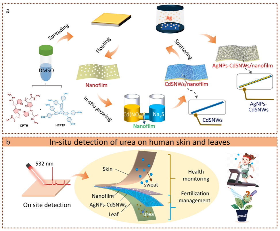
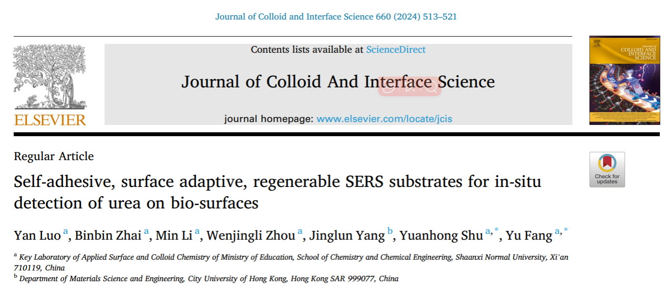
Figure 1. (a) Schematic illustration of the structure and application of SERS substrate in this work. (b) The preparation of the SERS substrate, AgNPs-CdSNWs/nanofilm.
Figure 2. (a) Optical photographs, (b) UV-vis spectra and (c-e) SEM images of the nanofilm, CdS NWs/nanofilm, and AgNPs-CdSNWs/nanofilm. (f) AFM images, (g) HR-TEM images and corresponding electron diffraction pattern, (h) EDS elemental mappings, (i) XPS spectra of AgNPs-CdSNWs/nanofilm. (j) The PXRD pattern of CdSNWs/nanofilm.

Figure 3 (a) Raman spectra of R6G, respectively, on silicon, AgNPs-silicon, nanofilm, AgNPs-nanofilm, CdSNWs/nanofilm and AgNPs-CdSNWs/nanofilm under the excitation laser of 532 nm. (b) Raman spectra of R6G molecules on the SERS substrate obtained at different R6G concentrations at an excitation wavelength 532 nm after baseline subtraction. (c) Intensities of the Raman peaks at 612 cm-1 at different R6G concentrations, with standard deviations as error bars. (d) SERS mappings of the signal intensity at 612 cm-1 for R6G. (e) Raman spectra of 10-5 M R6G after light irradiation at different times. (f) Reproducibility test for the SERS substrate upon 3 cycles of dropping 10-5 M R6G after light irradiation.
First Author: Luo Yan, doctoral candidate, Shaanxi Normal University
Correspondence Authors: Prof. Fang Yu and Dr. Shu Yuanhong, Shaanxi Normal University
Full Text Link: https://doi.org/10.1016/j.jcis.2024.01.068